
Advanced Microscopy Facility
At Wesleyan, we offer cutting-edge scientific imaging solutions to advance your research projects. Our state-of-the-art equipment includes the Leica SP8 conformal microscope and the HITACHI SU5000 Field Emission Scanning Electron Microscope (with energy-dispersible x-ray spectroscopy).
Our focuses are on biological and non-biological material analysis. At Wesleyan we serve a wide range of researchers from several departments, including; Biology, Molecular Biology and Biochemistry, Chemistry, Molecular BioPhysics, Earth and Environmental Sciences and others.
1 - Equipments (Technical Specifications):
Leica SP8 Confocal Microscope:
- Super resolution high speed imaging
- Provides exceptional imaging capabilities, allowing you to explore biological materials with high-resolution, 3D visualization
- Simultaneous multicolor imaging with true super-resolution (as fine as 120nm)
- Versatile features enable precise fluorescence and spectral imaging, making it ideal for a wide range of applications
- Leica's exclusive detection concept of LIGHTNING allows you to trace the dynamics of multiple molecules, even those expressed at low levels, simultaneously over long recording times in living specimens
Specifications:
- DMi8 inverted microscope stand (DMi8CEL)
- Super Z Galvo Stage; optional Universal or Multiwell plate inserts
- Transmitted light PMT
- Blue/green/red/far red filter cubes for wide-field fluorescence
- Lasers (two diode and 3 optically pumped semiconductor laser)
- Diode 405
- Diode 638
- OPSL 488
- OPSL 514
- OPSL 552
- Objectives:
- 5x (HC PL FLUOTAR 5x/0.15 DRY)
- Free working distance: 13.700mm
- 10x (HC PL APO CS2 10x/0.40 DRY)
- Free working distance: 2.740mm
- 20x (HC PL APO CS2 20x/0.75 DRY)
- Free working distance: 0.620mm
- 25x (water) (Fluotar VISIR 25x/0.95 WATER)
- Free working distance: 2.400mm
- 40x (oil) (HC PL APO CS2 40x/1.30 OIL)
- Free working distance: 0.240mm
- 63x (oil) (HC PL APO CS2 63x/1.40 OIL)
- Free working distance: 0.140mm
- 5x (HC PL FLUOTAR 5x/0.15 DRY)
- TCS SP8 Confocal Scan Head
- Non-resonant (conventional/variable) Scanner: 1 – 1800 Hz (7 fps @ 512 x 512 px, 84 fps @ 512 x 16 px), line frequency up to 3600 lines/second (bidirectional), max. scan format 8192 x 8192 px, scan field 22 mm diag.
- Resonant Scanner (ultra-fast, reduced photo damage): 8000 Hz (28 fps @ 512 x 512 px, 290 fps @ 512 x 16 px), line frequency up to 16,000 lines/second (bidirectional), max. scan format 2496 x 2496 px, scan field 13 mm diag. Optical field rotation 200°.
- Detectors: Lambda-Scan mode permits recording of spectral image series
- 2 HyD hybrid Spectral detectors:
- Very low dark noise
- Photon counting capability
- Enables time gated detection
- 1 PMTs:
- 40 MHz sampling
- Detection range 400–800 nm
- Transmitted light detector (TLD)
- Ideal for bright image acquisition
- 2 HyD hybrid Spectral detectors:
HITACHI SU5000 Scanning Electron Microscope (SEM):
- Ideal for characterizing surface structures, particles, and materials with utmost precision
- Suitable for conductive and non-conductive materials
- Ideal for variable pressure, allowing for high and low voltage image analysis
- Super high-resolution imaging at a nano scale level
Types of Detectors:
- Dual secondary electron (SE) detector
- Back Scatter detector (BSD)
- Ultra-variable Pressure Detector (UVD)
- Energy Dispersible X-ray Spectroscopy (EDS)
X-ray detection used to determine the elemental composition of materials. Each element produces characteristic X-ray peaks at specific energy levels.
Services Provided
- Training New Users
- Support during image acquisition
- Sample preparation of biological material for SEM analysis
Want to analyze your biological samples on the SEM but don't know where to start?
At the imaging facility you can get help preparing you smaples SEM imaging. expand... We can prepare (fix, dehydrate, critical point dry, mount and carbon coat)
Fess
Microscopes
- $X/hour - Expert user (fully trained)
- $X/hour -Users who require Dr. Pimenta's oversight
- $X/hour - Dr. Pimenta will operate the equipment and be responsable for all image acquisition
- $0 - Training on confocal or SEM
Samples Preparation for SEM
- $X/hour -
Steps For Biological Samples Preparation
Biological material requires special preparation before image analysis on the SEM. Here we outline the complicated steps necessary to preserve the surface morphology of most living tissue. Below is an overview of the process involved in sample preparation.
- Fixation
- Post-fixation
- Dehydration
- Critical Point Dry
- Mounting
- Gold Sputter
1) Fixation: In scanning electron microscopy, biological samples are fixed and prepared to withstand the vacuum conditions and electron beam exposure. The most common fixative agent for biological samples in SEM is glutaraldehyde. At Wesleyan’s imaging facility, we have adapted a protocol where we use 5% Glutaraldehyde in 0.1M Sodium Cacodylate Buffer to fix our biological material. Glutaraldehyde is a bifunctional aldehyde that cross-links proteins by forming covalent bonds between amino groups. This cross-linking stabilizes the cellular structures, preventing them from degrading during the SEM preparation process.
- Characteristics: Preserves cellular morphology by maintaining the integrity of cell membranes and structures, it also minimizes shrinkage of biological samples, accurately representing the original size and shape of cellular components. This fixative also has ideal penetration allowing it to quickly diffuse through tissues and cells. This ensures that the fixative reaches all parts of the sample for uniform fixation.
2) Post-fixation with 1% Osmium tetroxide and 0.8% Potassium Ferricyanide: Osmium tetroxide is a heavy metal stain that enhances the electron density of cellular structures. The addition of potassium ferricyanide to osmium tetroxide can introduce selective staining effects. Potassium ferricyanide reacts with osmium tetroxide to form osmiophilic ferricyanide precipitates, which selectively stain certain cellular components. This can provide additional information about the composition and distribution of specific structures within the sample.
- Characteristics: osmiophilic ferricyanide has an affinity for membrane structures. This is useful for highlighting and preserving the details of cell membranes. Post-fixation can also help reduce the formation of artifacts that may occur during the fixation process. For example, it may minimize distortion or shrinkage of cellular structures, and ultimately helps with the preservation of the ultrastructure of cellular components.
3) Dehydration: Dehydration is a crucial step in the processing of samples for scanning electron microscopy. The primary reason for dehydrating samples is to replace water found in all biological samples. The sample are subjected to a series of acetone washes of increasing concentration (stating at 30% until 100%). This series of washes gradually removes water from the sample, and substitute the water with acetone. It's important to dehydrate the sample gradually to avoid rapid changes that could cause structural damage.
Dehydrating samples ensures:- Vacuum compatibility: Under high vacuum (a necessary condition for SEM), water vapor can cause electron scattering, leading to a decrease in image resolution and quality.
- Less Charging effects: Water is a poor conductor of electricity, and the presence of water on the sample surface can lead to charging effects when exposed to the electron beam. Dehydration helps in reducing or eliminating charging effects, ensuring that the SEM images accurately represent the surface morphology of the sample.
- Improved resolution: Dehydration enhances the electron transparency of the sample, allowing electrons to penetrate the sample more effectively. This results in improved imaging resolution and detail, as the electron beam can interact more efficiently with the specimen.
- Preservation of Sample Structure: Dehydrating the sample helps to preserve its structural integrity. Water, especially in cellular structures, can cause distortion and shrinkage during the drying process. Removing water and replacing it with a dehydrating agent helps maintain the original size and shape of the sample.
4) Critical Point Drying: After dehydration, samples are immediately dried by a process known as critical point drying. This helps to avoid the surface tension-related damage that can occur during traditional air-drying or other drying methods. This is based on the principle of transitioning liquid CO2 to a gas without passing through the liquid-gas interface. At the critical point, the supercritical fluid exhibits properties of both liquids and gases. It has the density of a liquid but the diffusivity of a gas. Importantly, there is no distinct interface between the liquid and gas phases. Due to the absence of a liquid-gas interface, critical point drying allows for a gentle transition from the transitional fluid to the gas phase. Critical point drying minimizes or eliminates the artifacts associated with conventional drying methods, such as shrinkage, collapse, or distortion of delicate biological structures.
- The steps involved in critical point drying include:
- The dehydrated samples in 100% acetone are transferred immediately into a critical point dryer machine
- Liquid CO2 is introduced into the chamber and under pressure the acetone is slowly expelled and liquid CO2 takes it place.
- The temperature inside the pressurized chamber is slowly increased and the CO2 undergoes a phase transition, also known as critical point.
- In this point crucial step, the transitional fluid exists as neither a liquid nor a gas but as a supercritical fluid. For carbon dioxide, the critical point occurs at approximately 31.1°C (88°F) and 73.8 atmospheres of pressure.
- Once the sample has been dried using the supercritical fluid, the pressure is slowly released, allowing the carbon dioxide to revert to a gas. The gas is then vented from the pressure vessel, leaving behind the dried sample.
5) Specimen Mounting: After critical point drying, the samples are carefully mounted on special metal supports known as stubs. For this, we use a conductive double-sided carbon tape and one side is fixed to the stub and the other side is where the samples is carefully placed. The conductive tape helps to provide good electrical contact between the sample and the stub, minimizing charging effects during SEM imaging. It is important to note that after critical point drying, the samples are very brittle and can easily break.
6) Gold Sputtering: The final step before loading the samples into the SEM involves the use of a gold sputter machine. Gold sputtering involves depositing a thin layer of gold onto the sample surface, which serves multiple purposes, including enhancing conductivity, reducing charging effects, and improving the overall image quality. It is typically carried out using a technique called "sputter coating." Sputtering is a physical vapor deposition (PVD) process where energetic ions bombard a target material, causing atoms to be ejected from the target and depositing onto a substrate. This process occurs under a vacuum and in the presence of an inert gas, typically Argon. Argon is commonly used as it is inert and readily forms a plasma when subjected to a high voltage. Other types of metal can also be used but for most application gold is the most commonly used metal.
- Gold sputtering steps:
- Load dried and mounted samples in the machine.
- The chamber is pressurized, typically in the range of 10^-2 to 10^-5 torr. This low-pressure condition is necessary for the sputtering process to occur efficiently.
- Argon gas is introduced into the vacuum chamber. Argon is commonly used as it is inert and readily forms a plasma when subjected to a high voltage.
- A high voltage is applied to the argon gas, leading to the ionization of argon atoms and the formation of a plasma.
- The positively charged argon ions in the plasma are accelerated towards the gold target. Upon collision with the target, gold atoms are dislodged and are deposited onto the sample surface, forming a thin coating. Thickness can be adjusted by controlling sputtering time.
- The thickness of gold sputtering for biological samples in scanning electron microscopy (SEM) is typically in the range of 2 to 10 nanometers.
-
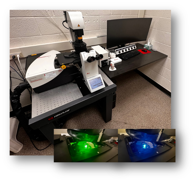
Leica Confocal
-
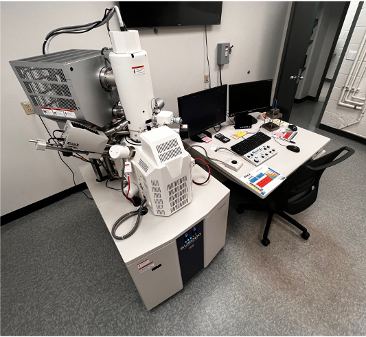
SEM
-
-